
ImmunitasBio Enzyme-Linked ImmunoSpot (ELISpot) platform provides robust data on cell-mediated immunity for vaccines and onco-immune targets. As the antigen-specific cells are activated, they release the cytokine of interest, which is captured directly on the membrane surface by the immobilized antibody. The visualization of the cytokine secreted by individual T- or B-clones is achieved by enzyme-conjugated or fluorescence-based substrates. A fluorescence-based method allows multiplex analysis of antigen-specific clones. The spots are then counted with the automated ELISpot reader system.
The ELISpot platform is considered to be one of the most useful for determining cell-mediated immunity because of its sensitivity and specificity in detecting rare antigen-specific T-cells (or B-cells). ELISpot assays are able to provide qualitative information regarding the specific cytokine or other secreted immune molecules.
We use ELISpot to visualize the secretory product(s) of individual activated or responding cells to analyze the frequencies of rare antigen-specific cells within a test population, such as single positive cells within a population of peripheral blood mononuclear cells (PBMCs).
ImmunitasBio utilizes ELISpot in our work with Cytotoxic T-Cell Response, Helper T-Cell Response, B-Cell Antibody response, Epitope Discovery. Specific to the bioanalytical assessment of cell and gene therapies, ELISpot can be used to assess cellular immune responses against the vector.
How ELISpot Works:
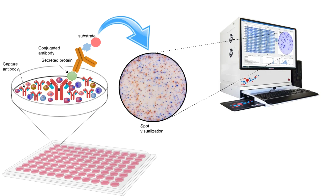
The enzyme-linked ImmunoSpot (ELISpot) assay is a highly sensitive immunoassay that measures the frequency of cytokine-secreting cells at the single-cell level. In this assay, cells are cultured in a 96-cell plate with a special membrane coated with a specific capture antibody. In the presence of a specific stimulus, proteins, such as cytokines, are secreted by the cells and captured by specific antibodies. After an appropriate incubation time, cells are removed and the secreted molecule is detected using a detection antibody in a similar procedure to that employed by a sandwich ELISA. The detection antibody is either biotinylated and followed by a streptavidin-enzyme conjugate or directly conjugated to an enzyme. By using a substrate that precipitates, the end result is visible as spots on the surface of the membrane. Each spot corresponds to an individual cytokine-secreting cell.