Biomarkers via Advanced IHC & Spatial Imaging
- Drug Discovery & Preclinical
- Monoclonal Antibodies
- Antibody-Drug Conjugates
- Vaccines (Prophylactic & Therapeutic)
- Cell & Gene Therapies (CGT)
- Allergy & Hypersensitivity
- Biomarker
- High-Dimensional Single-Cell Analysis by Flow Cytometry
- High-Throughput Cytokine & Chemokine Profiling by Multiplexing
- Biomarkers via Advanced IHC & Spatial Imaging
- Rare Cellular Functional Biomarkers by ELISPOT
- Real-Time Kinetic Biomarkers by IncuCyte S3
- Genomic Biomarker Analysis by qPCR & Droplet Digital PCR
- Seahorse XF: Immunometabolism & Bioenergetics
- MALDI Biotyper: Rapid Microbial & Proteomic Profiling
- Sapidyne KinExA
- Robust Protein Quantification by ELISA
- Advanced LNP Formulation & Screening – Unchained Labs Sunshine
- Translational & Clinical Research
- Immunogenicity Risk Assessment & Immunotoxicology
- Regulated Bioanalysis: PK, PD & Soluble Biomarkers
- Histopathology & Spatial Biology
- Advanced Flow Cytometry & Cellular Immunology
- Molecular Bioanalysis & Genomic Profiling
- Functional Potency & Ex Vivo Translational Models
- Central Laboratory & Clinical Trial Sample Management
- Dermo-Cosmetics & FMCG
- Nutraceuticals & Wellness
- Biorepository
Biomarkers via Advanced IHC & Spatial Imaging
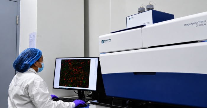
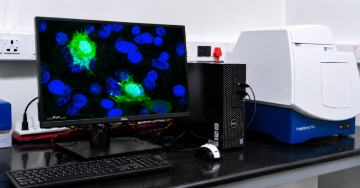
Understanding the localized impact of a therapeutic requires preserving the complex architecture of intact tissue. By integrating highly multiplexed Immunohistochemistry (IHC) with advanced spatial imaging, we provide a continuous analytical thread from early drug discovery in animal models to definitive biomarker evaluation in human clinical trials.
This high-dimensional spatial data accelerates decision-making across the entire translational pipeline:
Biodistribution & Target Engagement:
Visually map exactly where targeted biologics, advanced cell therapies, or ternary polyplex formulations localize within animal tissues. This confirms target engagement at the single-cell level and strictly validates the mechanism of action (MoA) in vivo.
Preclinical Spatial Toxicology:
Detect localized, tissue-specific toxicity or off-target binding early in development. By identifying exactly which cell populations are adversely affected in preclinical models, teams can confidently de-risk candidates before IND submission.
Microenvironment Characterization:
In complex disease models such as syngeneic tumor models or induced autoimmune tissues, spatial profiling quantifies immune cell infiltration, cell-to-cell interactions, and the reorganization of the structural stroma in response to treatment.